New Imaging Technology May Help in Diagnosing Neuropsychiatric SLE, Study Suggests
Written by |
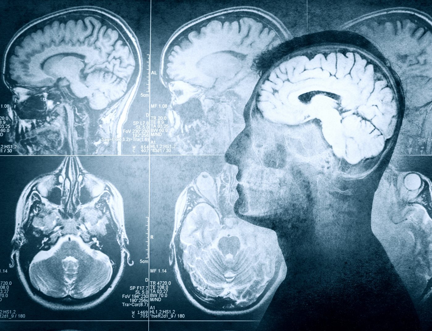
A new type of functional magnetic resonance imaging — called 3D arterial spin labeling (3D-ASL) — that measures blood flow in the brain may help in the early detection of lupus affecting the brain, a study suggests.
The study, “Cerebral blood flow abnormalities in neuropsychiatric systemic lupus erythematosus,” was published in the journal Lupus.
When systemic lupus erythematosus (SLE) affects the brain, it’s called neuropsychiatric SLE (NPSLE). This condition is hard to diagnose.
No consensus exists on what features are needed for a diagnosis of NPSLE, and cognitive changes indicative of the condition can be easily written off or ignored by all.
This presents a major problem because once the brain is damaged, it’s virtually impossible to have any meaningful healing; early detection, with treatment aimed at preventing damage before it occurs, is crucial.
Researchers hypothesized that 3D-ASL might be a viable way of detecting NPSLE lesions in brain scans, potentially improving diagnostics. This imaging technique works by tracking water molecules, allowing measurement of cerebral blood flow (CBF) — basically, where there is blood flowing in the brain, with irregular flow indicating lesions.
They used 3D-ASL to measure the brains of 16 people with NPSLE, and 19 people with SLE but no neurological symptoms. Thirty people without autoimmune diseases were included as a control group; all participants were female.
All in the control group had “homogeneous and symmetric perfusion” in various brain areas, the researchers wrote. Essentially, there was no evidence of irregular blood flow in the non-SLE brains.
All those with lupus (both SLE and NPSLE) had at least some irregular blood flow. For instance, the average relative CBF value in the frontal lobe was 6.47 among lupus patients and 2.07 among the controls, a statistically significant difference; similar differences were found in other brain regions as well.
NPSLE patients were more likely to have brain lesions detected by 3D-ASL. Depending on the specific area of the brain under scrutiny, NPSLE patients accounted for between 60% and 100% of the detected lesions.
“Collectively, our results indicated that neurological lesions in SLE patients can be detected by ASL, and CBF is helpful for the early diagnosis of NPSLE,” the researchers concluded.
This study was a relatively small pilot study; further research will be needed to figure out exactly how this method can be employed for maximum reliability and efficacy.
Data already demonstrate that this method is not infallible. For instance, all frontal lobe lesions detected were in NPSLE patients, but not all NPSLE patients had frontal lobe lesions, so this isn’t a “one-and-done” diagnostic test. But it adds to the available diagnostic tools that may allow NPSLE to be diagnosed and treated earlier and, by extension, more effectively.