Inhibitor of Nogo-a Protein Improves Symptoms of Neuropsychiatric Lupus in Mice, Study Shows

A new study suggests that the protein Nogo-a, known to limit the recovery of neurons after injury, is involved in the development of neurophsychiatric lupus (NPSLE).
When this protein was inhibited in a mice model of the disease, mice had better myelin repair, improved cognitive and memory function, and lower levels of pro-inflammatory proteins, suggesting that this could be a possible therapeutic approach for lupus patients.
The study, “Neuropsychiatric involvement in lupus is associated with the Nogo-a/NgR1 pathway,” was published in the Journal of Neuroimmunology.
Approximately half of patients with systemic lupus erythematosus (SLE) exhibit neurological or psychiatric symptoms, collectively categorized as neuropsychiatric lupus (NPSLE). This form of the disease is particularly severe, with patients experiencing seizures, depression, anxiety, psychosis, and cognitive disorders.
While the causes underlying NPSLE are not fully known, neuroinflammation and neurodegeneration have been described as significant contributors.
The Nogo-a protein had been shown to limit recovery of the adult central nervous system after injury, which led a team of Chinese researchers to study the link between this protein and its receptor, NgR1, and the development of NPSLE.
Looking at cerebrospinal fluid, a protective body fluid found in the brain and spinal cord, the team found that Nogo-a was only present in patients with NPSLE, but not in patients with other autoimmune disorders.
“These results suggest that Nogo-a may be specifically expressed in patients with NPSLE and is likely associated with NPSLE pathogenesis,” the authors explained.
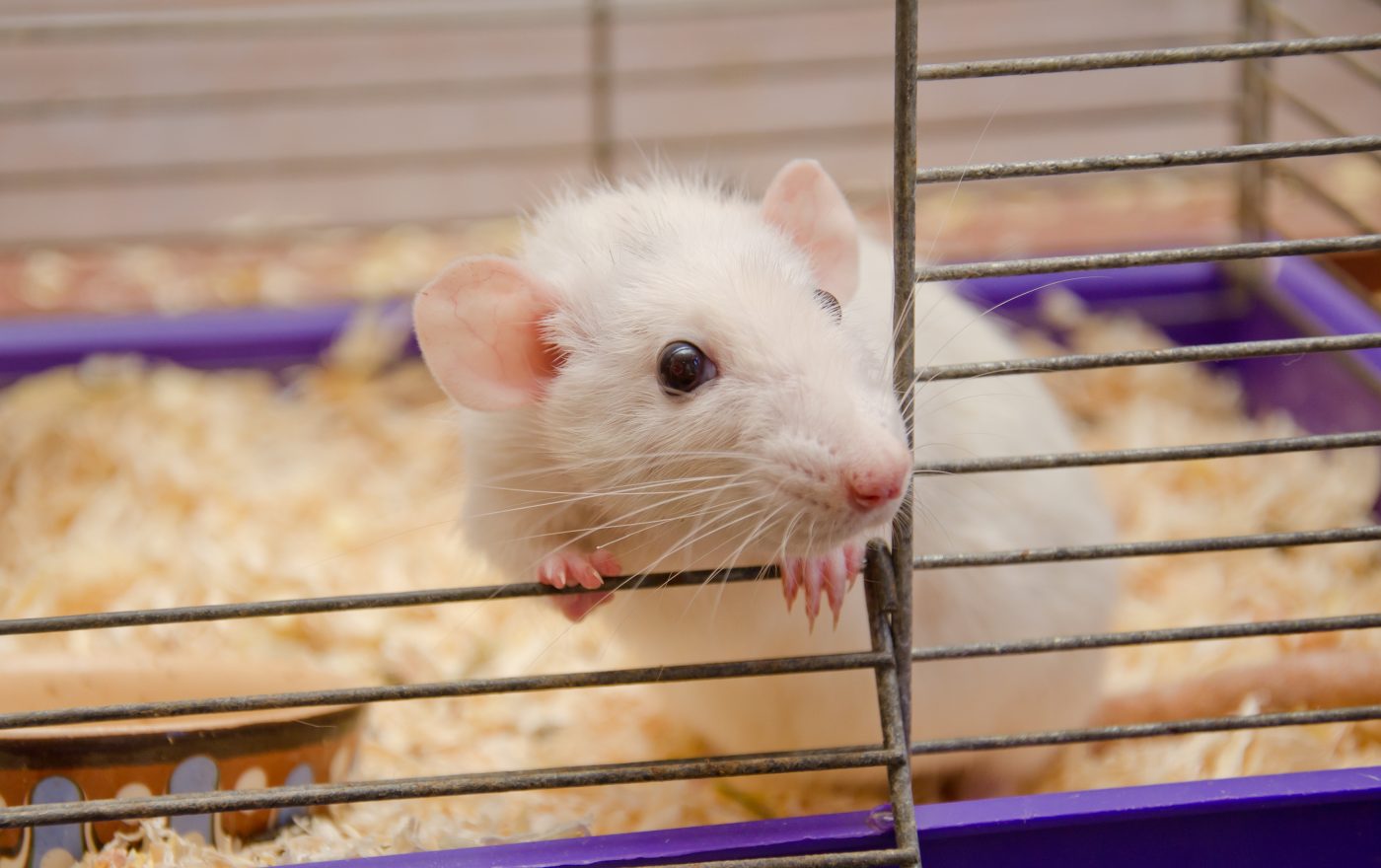
To further explore the role of Nogo-a in SLE, the team used an animal model of lupus that spontaneously develops symptoms typical of lupus and the cognitive and affective disorders characteristic of NPSLE.
As the mice aged, they showed increasing inflammation and nerve damage, a phenotype, the researchers found, that paralleled with increased levels of Nogo-a. In fact, NPSLE mice all had Nogo-a positive neurons, and exhibited destruction of their myelin sheath and increased levels of pro-inflammatory proteins in the brain.
“The Nogo-a protein might play a role in inflammation and nervous damage during aging in MRL/lpr mice,” the researchers said.
Mice treated with an inhibitor of Nogo-a, called NEP1-40, had reduced neuronal damage and decreased neuroinflammation compared to non-treated mice.
“This study highlighted that Nogo/NgR signaling might be involved in the pathogenesis of NPSLE and that functionally [inhibiting] Nogo-a could be used as a potential target for the treatment of neuropsychiatric lupus,” the team concluded.


