Lupus Researchers Reveal Potential New Approach to Controlling Inflammation, and Tissue Damage
Written by |

Researchers from St. Jude Children’s Research Hospital in Memphis, Tennessee, in collaboration with several other institutions, published findings from a lupus study in which they discovered a new approach to controlling the inflammation and tissue damage that is a hallmark of the chronic autoimmune disorder.
The study, “Noncanonical autophagy inhibits the autoinflammatory, lupus-like response to dying cells,” was published in the latest edition of the journal Nature.
The discovery was made by Jennifer Martinez, Ph.D., first author and a postdoctoral fellow in the laboratory of senior researcher Dr. Douglas Green, Ph.D., lead study author and chair of the Department of Immunology. Martinez was the first to notice that mice with a defect in a process for digesting dead cells called LC3-associated phagocytosis (LAP) were significantly smaller than related mice without the defect.
This observation made her question if LAP, an important immunological component that makes certain the dead and dying cells are digested and disposed of properly after being eaten by immune scavenger cells called macrophages, are involved in the systemic lupus erythematosus (SLE) disease process.
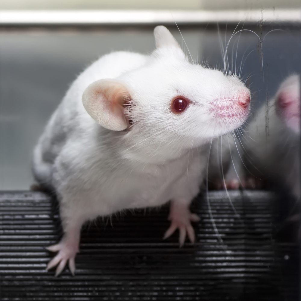
To answer this question, Martinez and colleagues used experimental mice lacking any of the several components of the LAP and mice that were sufficient in LAP, and injected them with dying cells to assess the difference in the levels of inflammation, and inflammatory mediators, as well as kidney function, between the two groups of animals.
The findings showed that:
- The mice lacking any of several components of the LAP pathway show increased serum levels of inflammatory cytokines and autoantibodies, glomerular immune complex deposition, and evidence of kidney damage.
- When dying cells were injected into LAP-deficient mice, they are engulfed but not efficiently degraded and trigger acute elevation of pro-inflammatory cytokines.
- Repeated injection of dying cells into LAP-deficient, but not LAP-sufficient, mice accelerated the development of SLE-like disease, including increased serum levels of autoantibodies.
“We hope the findings offer a window into the cause of this devastating disease in some patients and an opportunity to improve treatment by developing novel approaches to prevent or reduce the inflammation and autoimmune response that characterize lupus,” Green said in a press release.
“Defects in the clearance of dying cells have been implicated in the SLE disease process, and previous studies have suggested that the problem may stem from defects in autophagy. In this study, we showed that LAP contributes to SLE in mice and likely accounts for the previously reported genetic association between autophagy and lupus,” Green said.


