
Backdoor Way of Regulating T-cells Seen to Lower Kidney Inflammation, Damage in Lupus Mouse Model


Researchers found that controlling the activity of Tbet, the main transcription factor of inflammatory T-cells, reduces kidney inflammation and tissue damage in mice prone to develop lupus.
The study, “Intranuclear delivery of the transcription modulation domain of Tbet-improved lupus nephritis in (NZB/NZW) F1 lupus-prone mice,” was published in Kidney International.
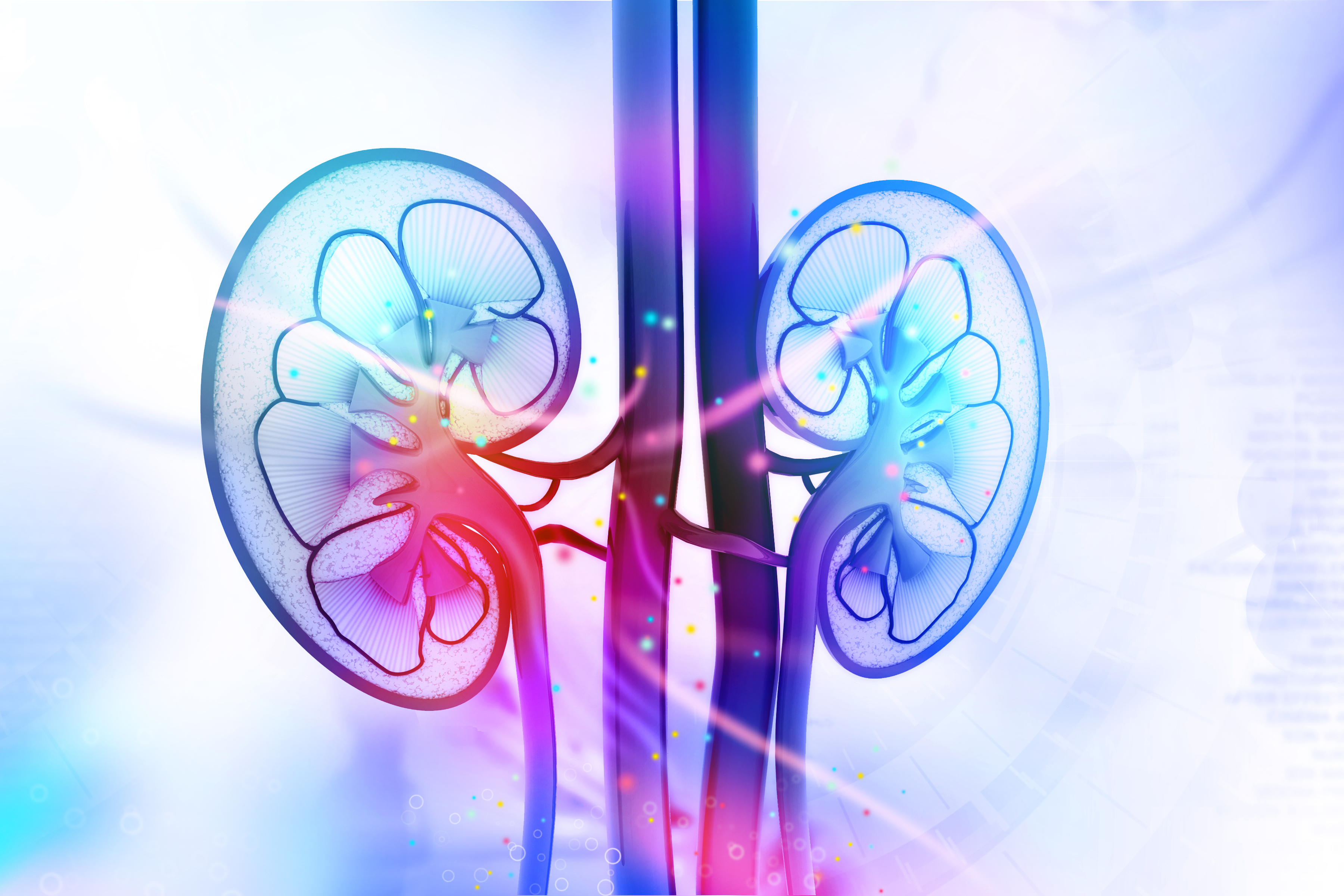
Lupus nephritis (LN) — inflammation of the kidneys caused by lupus — can be common in systemic lupus erythematosus (SLE) patients. It is characterized by an excess of proteins in the urine (proteinuria) and physical alterations in kidney tissue.
Recent studies suggest that inflammatory T-cells, which are essential for a healthy immune response, aggravate LN by boosting local tissue inflammation.
Monoclonal antibodies that target and control the activity of T-cells are now considered the main therapeutic agents for a number of autoimmune diseases. But they are less effective than steroid-based injections and oral immunosuppressants, which are the standard treatments for LN.
These standard therapies, however, have major side effects, including lowering the number and activity of T-cells. This posed a serious question to researchers: How can T-cell activity be effectively fine-tuned without compromising the immune response?
The researchers in this study developed a new way of controlling T-cell activity — that of blocking Tbet, a transcription factor that controls the expression of genes involved in a cell’s response to infection.
To inhibit Tbet function, they delivered the transcription modulation domain (TMD) of Tbet directly into the nucleus of cells grown in the lab and those from animal models of disease.
By delivering this form of TMD (called ntTbet-TMD), the researchers managed to prevent the production of inflammatory substances that lead to LN onset and progression.
The studies performed in mice prone to lupus (NZB/NZW model) showed that proteinuria decreased by 58.1% and 73.8%, respectively, in animals given low and high doses of ntTbet-TMD compared to an untreated group.
Mice from the untreated group also had extensive damage to kidney tissue — expanded glomerulus (the filtering units in the kidneys), high cell proliferation, and inflammatory cell infiltration — that was milder in animals treated with ntTbet-TMD.
No side effects were reported in the ntTbet-TMD group.
“Importantly, in contrast to methylprednisolone and immunosuppressive agents, ntTbet-TMD can be a novel and highly specific immunotherapy for the treatment of LN without affecting the differentiation of other T-cell subsets and T-cell activation events,” said the investigators regarding the therapeutic potential of ntTbet-TMD compared to conventional therapies.
“This fundamental technology to deliver the wild-type or dominant negative form of the transcription factor into the nucleus of the cells in vitro and in vivo can be a core technology in developing novel therapeutics for various autoimmune diseases in which specific transcription factors play a key role in the pathogenesis,” they concluded.